Five years ago the researcher Barbora Špačková moved to Sweden to take up a postdoc position at the Chalmers University of Technology where she developed a unique imaging method called nanofluidic scattering microscopy using which she can see, as she puts it, “things that nobody else has ever seen before”. Putting the method at the heart of her business plan, she established a spin-off company Envue manufacturing microscopes used for single-molecule imaging. Recently she has returned home to the Czech Republic, settling down at the Department of Optical and Biophysical Systems of the Optics Division of the Institute of Physics, where she would like to set up her own research team. Her article on nanofluidic scattering microscopy has lately been published by the prestigious Nature Methods.
What exactly is the nanofluidic scattering microscopy method built on?
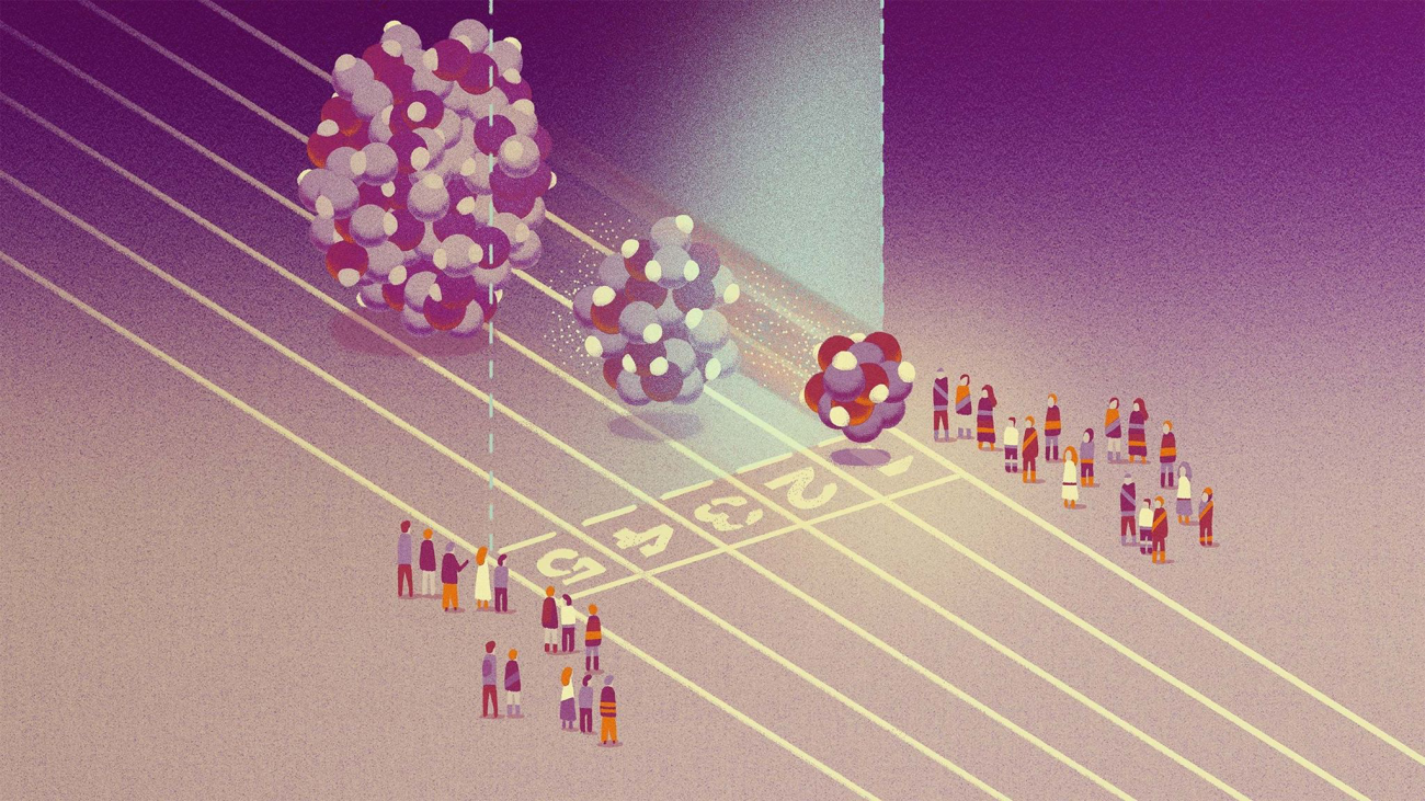
All objects scatter light, which is why we can observe, for example, even very small dust particles. But biomolecules are by several orders of magnitude smaller. They do scatter light but only a little, which makes it impossible to detect it. The trick I use to detect such scattered light is based on the fact that light behaves as a wave, and once you let it interfere with some other light, you can make a small intensity become a higher one. In other words, if I shine a light on a nanochannel which contains a biomolecule, this nanochannel scatters light too, and its intensity, compared to the scattered light on the biomolecule, is huge. The light scattered by the biomolecule then interferes with the light scattered by the biochannel. And that’s where I can see things that nobody else has seen before.
What is the main benefit of this method?
I am a physicist – to me biomolecules are just like little marbles, and I like to overstep the boundaries of the possible. But for researchers concerned with the study of biological processes this means that they can use this method to study individual biomolecules in their natural environment and in their natural motion, which was impossible in the past. The traditional method used to observe single molecules is fluorescence microscopy, which has been used for decades. It uses some kind of “beacon” – a fluorescence marker, emitting light, which you place on a molecule. Then you don’t observe the target molecule but the marker. However, in many cases, it turned out that the marker influences the molecule. But if you want to study life in its natural environment, you want to set as realistic conditions as possible. A ground-breaking optical method is the iSCAT, for example; this can capture molecules even without using the markers but they need to be attached to some kind of support or an object. Not all molecular processes take place on a surface. It often happens to me that when I show how molecules move around in an aqueous environment to a biochemist or biologist, he or she experiences a vow effect – they finally see how a molecule behaves naturally.
So, you place emphasis on the natural side of things in your research…
Yes, and not only on it. Using this method we can also conduct a real-time study of biological processes, and to measure the dimensions and weight of individual molecules.
Have you compared fluorescence microscopy with your method? Is there such a big difference? Does the marker influence the molecule significantly?
We’ve done one experiment related to DNA, in which we observed enormous differences – this research is still in progress – but these enormous differences are no general conclusion that would be applicable to other molecular processes.
How did you come up with the concept of nanofluidic scattering microscopy and what preceded it?
At Chalmers I took up a postdoc position, and I must say that initially I had a lot of freedom in what I wanted to do. This was a great situation for someone like me. There were lots of people around me with huge know-how and profound knowledge to which I hadn’t had access before. So, during the first few months I moved around nanofluidics, which was a completely new field to me, exploring its boundaries and the ways people made use of it…
It looks like it captured your attention right from the very beginning….
That’s right. I was thinking about how I could combine this field, which was a completely new one to me, with my research focus – the application of optical methods to biochemistry, and, at a more general level, to biosciences. Actually, it all started with a conversation with my colleague. He showed me a strange effect that didn’t work for him and that he wanted to get rid of, but he had no idea what caused it. I thought it was interesting and I started to formulate a theory which describes the interaction of light with nanofluidics because nobody else had studied this yet. The theory successfully explained what the problem was, while showing an incredible potential for microscopy. My conclusions showed that we can observe single molecules moving within a nanochannel.
This sounds almost incredible to me: you try to help a colleague while finding your feet at a new job, and you end up making a major discovery…
Frankly, the initial version of my theory was quite counterintuitive, and its result seemed too incredible. When I consulted the result with others at the university, I was often faced with doubts on the part of both the professors and my colleagues. Few people believed that something like that would be possible.
This might have been a bit discouraging for a young researcher, right?
My supervisor at that time Prof. Christoph Langhammer saw a great potential in the theory and was giving me a lot of support. Together we started building an apparatus, and it didn’t take us long – perhaps a half a year – to make the first real observation of biomolecules. I must admit that it was an absolutely great feeling to see my idea, which was first just numbers and equations on my blackboard, eventually translate into reality.
At the moment, you and your colleagues from the Chalmers University of Technology have published an article in Nature Methods – what exactly does it describe?
In the article we discuss not only how to observe biomolecules but also how to sensitively weight them a determine their dimensions; they are parameters of importance with, for example, proteins. We also describe our analysis of detection of exosomes, which are bodies produced by cells in living organisms. Up until recently, little was known about them, but lately their research has seen a huge boom. In principle, exosomes can be called cell “minicopies” that contain a lot of information. Once we are able to collect information about them, we will be able to study how the cells are doing, and detect, for example, different diseases. The characterisation of such particles is a hard job because each of them is slightly different, contains a different piece of information, they are rather heterogeneous. Therefore, many researchers tend to choose methods that can do particle-after-particle characterisation so they can read the information correctly.
…to be able to use it for disease diagnostics?
Yes, that’s the plan. At the moment, we are at the proof of concept phase, at which researchers want to find out what structure and composition exosomes have, how heterogeneous they are, and which of them are of importance.
Now let’s talk about your spin-off company Envue Technologies, which was founded by you and your colleagues on the basis of your discovery. In it, you are developing microscopes based on the nanofluidic scattering microscopy method. Have you built one already, and if yes, is it on sale yet?
Currently we are developing a platform, which you might call a product. It has attracted interest from pharma companies or, generally, from researchers. This is a process that we expect to take several years. Anyway, now we have a laboratory prototype, apparatus I developed while I was at Chalmers.
How do you assemble a new microscope? Do you buy components and put them together?
The development of a laboratory prototype resembles a Lego set. There are sets available for developing optical systems. The laboratory prototype I built at Chalmers was based on some tailor-made devices, but it also consisted of many “Lego parts”. The end- product as such is an engineering solution and is much more robust.
Did you like playing with Lego as a child?
I still like playing with it. I have a child that, luckily for me, likes to play with it too, so, you might say, I build things at home, and I build things at the lab, though it’s not from Lego. In fact, a lot of scientific disciplines often remind me of childhood play sets.
Now let’s move on from research to talking more about you. After the years in Sweden, what made you decide to return to the Czechia?
At my career stage, people usually stand at a crossroads where they must decide if they see themselves more in academia or more industry, and whether they and their families want to live at home or abroad. This decision-making was very intense for me because I gravitate toward academia but I also established a company in Sweden. It was a hard nut to crack in many ways. We spent 4 and a half years in Sweden, which is an indelible part of our lives… If only you could see the whiteboard at home full of the pros and cons of staying or leaving to help us decide! We kept weighing up the pros and cons, until, eventually, we decided to go when I was awarded a GAČR grant. We packed things and left for Czechia within a month.
Why exactly did you choose the Optics Division and the Department of Optical and Biophysical Systems?
I have worked with Hanka Lísalová in the past, we stayed in touch, and it was her who gave me the idea. The Optics Division has grown considerably in the past two years, and it grew to include people I used worked with in the past. I collaborated Jakub Dostálek in a European project a few years ago, I have published several publications together with Scott Lynn, so returning back home was joining an environment I know well, with people I like to collaborate with. Also, the Division management has supported me and everything went smoothly there.
What is your vision for the FZU?
I have received two grants aimed to develop my nanofluidic scattering microscopy method in different directions. The GAČR grant is aimed at developing the method toward application for studying biomolecule interactions. The second one, the Marie-Sklodowska Curie Actions, is aimed at developing an important component – a nanofluidic valve using which I can improve the way I manipulate molecules. In the future, I’d like to set up a team to deal with the development of new single molecule tools so that scientists can be looking into the nano-world to unravel the mysteries of life at the molecular level.